
Research
Doctors May Now See the Hidden Signs of Sudden Cardiac Death
Columbia researchers have developed a new way to visualize the heart’s electromechanical activity, offering new insights into Mitral Valve Disease and arrhythmia risk.
Each year, roughly 2 to 5% of the population is affected by Mitral Valve Prolapse (MVP) and Mitral Regurgitation (MR). These conditions can affect the heart’s rhythm and have been linked to sudden cardiac deaths, particularly in advanced stages of disease, through the development of abnormal electrical activity. MVP has been found to bend the valve backward as the heart contracts, while MR affects the valve’s ability to close fully, leading to blood leaking backward.
Years after the development of Electromechanical Wave Imaging (EWI), Elisa Konofagou, the Robert and Margaret Hariri Professor of Biomedical Engineering at Columbia Engineering, has been developing techniques to capture electrical activity and mechanical motion within the heart based on cardiac ultrasound images called echocardiograms.
In a new study published today in the journal Proceedings of the National Academy of Sciences of the United States (PNAS), Konofagou and her team, in collaboration with Columbia NYP Pediatric Cardiology and the Institute of Cardiovascular Sciences in University College London, UK, found that patients with MVP had a significant electromechanically delayed activation of the left ventricle, particularly near the papillary muscles which ensure proper closure of the valve as the heart pumps blood. Subjects with even mild MVP and MR also noticed a longer full cardiac electromechanical activation and recovery time compared to healthy subjects. However, patients with MR experienced longer recovery time than those with MVP. The team successfully recruited patients with arrhythmogenic mitral valve disease, in whom disease progression had already resulted in ventricular arrhythmias.
“Our work has provided a new way to dive deeper into the important role that proper function of the mitral valve dictates how valve diseases affect the overall cardiac function and activation,” Konofagou said. “By using EWI to both detect and visualize these changes that are otherwise invisible by current clinical modalities, we hope to confirm it could be used for early detection and risk assessment, transforming how we evaluate and improve care for those suffering from mitral valve disease, but also arrhythmias in general.”

The team set out to determine whether EWI would be capable of identifying the changes in cardiac electromechanical activation and recovery in the pediatric population, where mitral valve disease could be isolated as a standalone comorbidity. The clinical study observed 21 pediatric subjects with healthy, MVP or MR affected hearts. Two adult arrhythmogenic MVP patients were also studied in the hopes of determining if the EWI could identify the source of the sporadic abnormal heartbeats alongside characterization of the electromechanical function in normal rhythm.
Using a single echocardiographic examination, the Konofagou team was able to identify and co-localize the source of the spontaneous arrhythmia with regions of locally delayed electromechanical function noninvasively. These findings were further corroborated by preclinical studies. This validation confirmed that the ultrasound technique can grant medical providers more critical information than the standard echocardiogram, providing diagnostic capabilities by looking at the heart as a complete system - electrical, mechanical, and valvular.
Currently, clinical echocardiograms are constrained to focus on the structure and mechanical function of the heart, such as how the mitral valve moves and how effectively the heart pumps the required blood volume at each heartbeat. The EWI builds on these capabilities to map how the electrical activation, and thereby electromechanical activity, moves perpetually through the heart. This added layer allows providers to identify subtle abnormalities that are not readily apparent through conventional imaging, whether by ultrasound, MRI, CT, or PET.
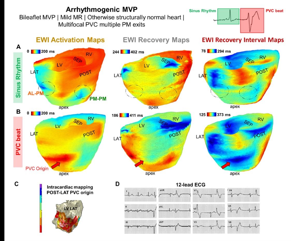
These findings indicate that the disease could be more than just a valve issue and that minute movements at very high framerates hold the key to divulging these important cardiac properties. The new question becomes, “Can these problems affect the timing and coordination of the surrounding heart muscle? And if so, how should we change the way we currently intervene?”
Looking to the future, the team will expand this testing to a larger population but also integrate the technology into clinical ultrasound scanners for everyday use at the point of care.
About the Study
Journal: PNAS
Authors: Melina Tournia, Christina Proestakia, Seungyeon Julia Hana, Johanna B. Tonkob, Mary Kucinskia, Rachel Webera, Rosalia Minyetya, Yaffa Wolickic, Youssef Elnabawid, Aikaterini Afentoulia, Jad El Harakea, Cagla Ozsoya, Hannah Schleifera, Leonardo Libermane, Alexandra Channinge, and Elisa E. Konofagoua