The 2024 Archimedes Dinner and Lecture Slideshow
The 2024 Archimedes Dinner and Lecture was held on November 20 at the Italian Academy


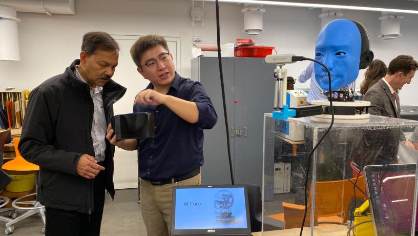
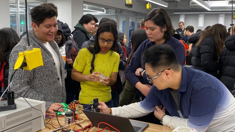
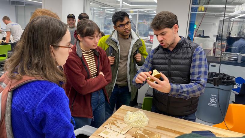
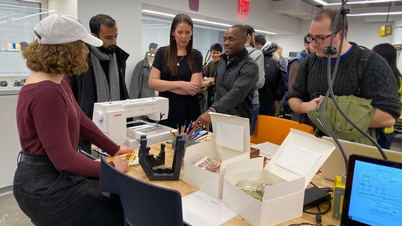
Columbia University's Makerspace
Explore the innovative hub for creativity and learning at Columbia University's Makerspace.
Mutations in the MYH9 Gene Cause Spectrum Disorders
Left: The fruit fly provides a unique opportunity to "watch" the effects of disease-associated mutations. Right: These changes in embryo shape require cell movements driven by mechanical forces.
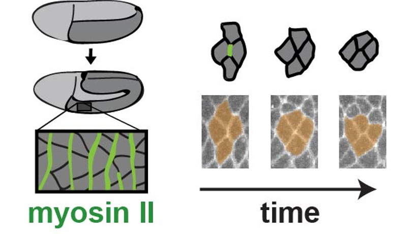
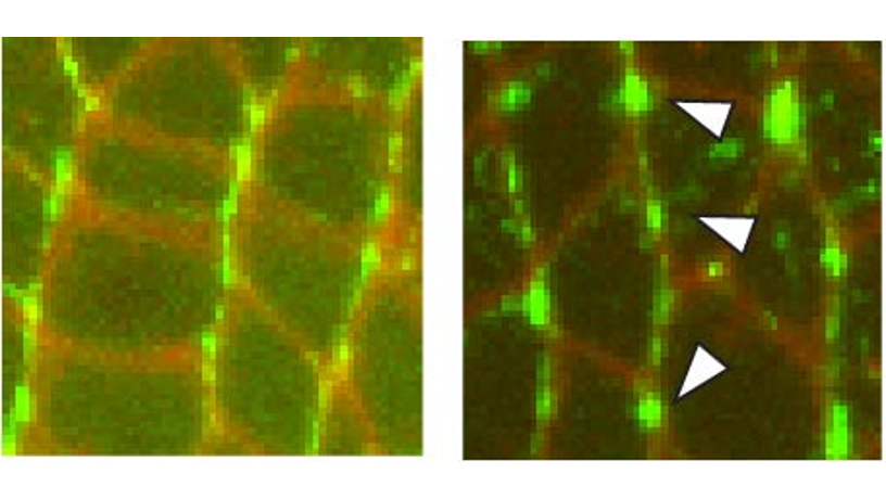
They used high-resolution confocal fluorescence imaging to take movies of the process, together with biophysical approaches such as laser ablation, or laser nano-dissection, to measure the forces generated by the mutated myosin II motor proteins in vivo.
Kasza found that, while the mutated myosin II motor proteins actually went to the proper places inside cells and were able to generate force, the fine-scale organization of the myosin proteins and the speed of their movement inside cells were different than for the normal wild-type myosin protein. The team saw slower movements of cells within tissues that brought about abnormalities in embryo shape during development.
“By ‘watching’ how cells move and generate forces inside living tissues, we’ve uncovered new clues as to why mutations in the MYH9 gene cause a broad spectrum of disorders in humans,” Kasza observes. “Our work sheds new light on how motor proteins generate forces inside living tissues and on how genetic factors alter these forces to result in disease. This mechanistic understanding will help us better understand these diseases and could lead to new diagnostic or therapeutic strategies down the road.”
The researchers are now working on new approaches to very precisely manipulate the forces generated by myosin motors inside living cells and tissues. These new tools will help the team to uncover how mechanical forces influence biochemical processes that control cell movements and cell fate. These studies will be essential to better understanding how dysregulation of mechanical forces contributes to disease.
Columbia Engineering
Columbia Engineering, based in New York City, is one of the top engineering schools in the U.S. and one of the oldest in the nation. Also known as The Fu Foundation School of Engineering and Applied Science, the School expands knowledge and advances technology through the pioneering research of its more than 220 faculty, while educating undergraduate and graduate students in a collaborative environment to become leaders informed by a firm foundation in engineering. The School’s faculty are at the center of the University’s cross-disciplinary research, contributing to the Data Science Institute, Earth Institute, Zuckerman Mind Brain Behavior Institute, Precision Medicine Initiative, and the Columbia Nano Initiative. Guided by its strategic vision, “Columbia Engineering for Humanity,” the School aims to translate ideas into innovations that foster a sustainable, healthy, secure, connected, and creative humanity.
Images by Karen Kasza/Columbia Engineering | Photo Credit: Karen Kasza/Columbia Engineering & Sara Supriyatno/Sloan Kettering Institute
About the Study
The study is titled “Cellular defects resulting from disease-related myosin II mutations in Drosophila.”
Authors are: Karen E. Kasza1,2,; Sara Supriyatno1; and Jennifer A. Zallen1.
1Howard Hughes Medical Institute and Developmental Biology Program, Sloan Kettering Institute;
2Department of Mechanical Engineering, Columbia Engineering.
The study was supported by NIH/NIGMS R01 grant GM102803 to JAZ. KEK holds a Career Award at the Scientific Interface from the Burroughs Wellcome Fund, a Clare Boothe Luce Professorship, and a Packard Fellowship. JAZ is an investigator of the Howard Hughes Medical Institute.
The authors declare no financial or other conflicts of interest.
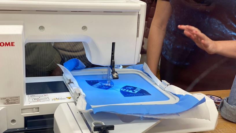
Robotically Assisted Preparation of a Microfluidic Diagnostic Chip
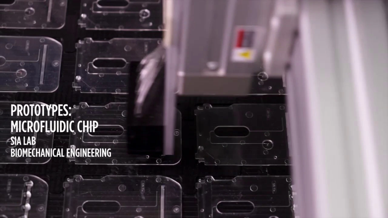
The researchers evaluated 142 samples, including patients with early Lyme disease, healthy individuals from areas where Lyme disease is endemic, and those with Lyme arthritis. They first screened a set of known diagnostic Lyme disease biomarkers for their ability to detect Lyme disease infection. They then tested the top three biomarkers using a standard enzyme immunoassay, and then mChip-LD, an advanced microfluidic platform developed by Sam Sia, to test the samples.
When tested against additional samples of serum from people with Lyme disease, the multiplexed set of biomarkers was more sensitive than standard Lyme disease tests, while also exhibiting high specificity. The team found that it was better at picking up signs of Lyme disease infection in early-stage samples—possibly because it was able to detect antibodies that peak in the first weeks after someone is infected with Lyme disease.
When the test was run on Sia’s mChip-LD platform, it worked very well, showing strong potential for the development of a point-of-care test for Lyme disease. “While the assay will require more refinement and testing before it can be approved for widespread use as a test for Lyme disease, our results are very exciting,” says one of the study’s lead authors, Siddarth Arumugam, who is a PhD student in Sia’s lab. “It will help so many people if we can develop a single, rapid, multiplexed diagnostic test to identify Lyme disease stage that can be used in doctors’ offices.”
Sia is the co-founder of Claros Diagnostics, whose underlying microfluidics technology is now being commercialized by OPKO Health and was recently approved by the FDA for testing for prostate cancer. He and Gomes-Solecki are now planning a more thorough clinical validation study to see whether the performance of the Lyme microfluidic platform holds up.
Columbia Engineering
Columbia Engineering, based in New York City, is one of the top engineering schools in the U.S. and one of the oldest in the nation. Also known as The Fu Foundation School of Engineering and Applied Science, the School expands knowledge and advances technology through the pioneering research of its more than 220 faculty, while educating undergraduate and graduate students in a collaborative environment to become leaders informed by a firm foundation in engineering. The School’s faculty are at the center of the University’s cross-disciplinary research, contributing to the Data Science Institute, Earth Institute, Zuckerman Mind Brain Behavior Institute, Precision Medicine Initiative, and the Columbia Nano Initiative. Guided by its strategic vision, “Columbia Engineering for Humanity,” the School aims to translate ideas into innovations that foster a sustainable, healthy, secure, connected, and creative humanity.
About the Study
The study is titled “A Multiplexed Serologic Test for Diagnosis of Lyme Disease for Point-of-Care Use.”
Authors are: Siddarth Arumugam a; Samiksha Nayak a; Taylor Williams b; Francesco Serra di Santa Maria a; Mariana Soares Guedes b,c; Rodrigo Cotrim Chaves a; Vincent Linder d; Adriana R. Marques e; Elisabeth J. Horn f; Susan J. Wong g; Samuel K. Sia a; and Maria Gomes-Solecki b,c.
a Department of Biomedical Engineering, Columbia Engineering
b Immuno Technologies Inc, Memphis, TN
c Department of Microbiology, Immunology and Biochemistry, University of Tennessee Health Science Center
d OPKO Diagnostics LLC, Woburn, MA
e Lyme Disease Studies Unit, Laboratory of Clinical Immunology and Microbiology, National Institute of Allergy and Infectious Diseases, National Institutes of Health
f Lyme Disease Biobank, Portland, OR
g Wadsworth Center, New York State Department of Health, Axelrod Institute
The study was supported by Public Health Service grant (R44 AI096551), and in part by the Intramural Research Program of the National Institute of Allergy and Infectious Diseases, National Institutes of Health.
Conflicts of interest: M.G.S. is an employee of Immuno Technologies, Inc, and V.L. was an employee of OPKO Diagnostics, LLC while engaged in the research project. We thank the National Institutes of Health, National Institute of Allergy and Infectious Diseases for funding support (grant R44 AI096551) to M.G.S. via Immuno Technologies, Inc. M.G.S. holds 5% or more financial interest in Immuno Technologies, Inc. M.G.S. and A.R.M. hold relevant patents. V.L. declares a financial interest in OPKO Health. S.A., S.N., F. S. S. M., T.W., R.C.C., M.G., S.J.W. and S.K.S. declare no competing financial interests.