Lead Photo Caption: Winter haze in Kolkata, India
Lead Photo Credit: Courtesy of V. Faye McNeill
The Lever: AI Agents at work

Credit: Jane Nisselson; Music: License agreement is made between Columbia Engineering and the copyright owner David Szesztay, in regards to the musical composition, titled as At the Right Moment.
About The Lever:
The Lever is a new collection of limited series newsletters direct from our faculty. In each edition, researchers at the forefront of discovery break a problem or technology into its core elements and explore the most likely pathways for progress. Over the course of a few days, subscribers will get an inside view into topics like sustainable energy, AI agents, or fusion energy.
Using Light to Study and Control Tissue Folding
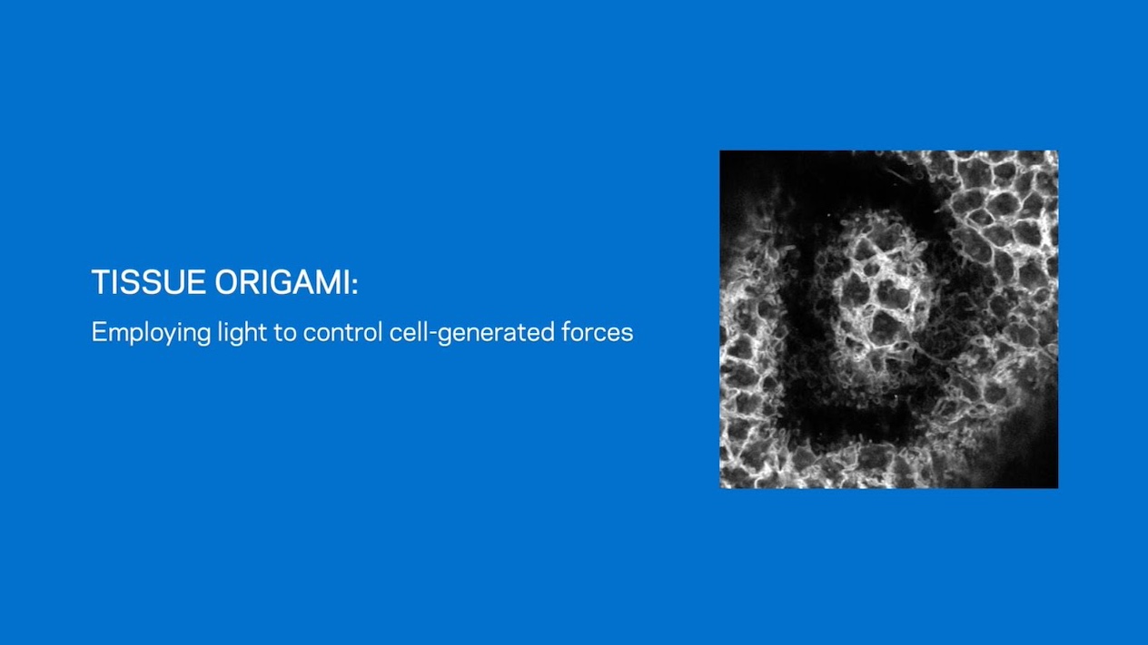
Opening video: an optogenetic tool illuminates a "D" shape in an early embryo tissue which causes the tissue to furrow in a "D" shape (D is for Drosophila melanogaster, the scientific name of the fruit fly).Following two videos: Optogenetic induction of tissue furrowing using two different optogenetic tools (OptoGEF2 and OptoCysts). Scale is 20 µm.
Video Credit: Andrew Countryman/Kasza lab
From flat sheets to complex structures
One major way that developing embryos build their organs is through furrowing — that is, they form pockets in tissues, which eventually become the sites of folds. "Just as a flat sheet of paper can be folded into a crane, a flat embryonic tissue can be folded into the precursor of an organ," said Andrew Countryman, a doctoral student in biomedical engineering at Columbia and the study's first author.
Previous research has developed many tools for manipulating the proteins and other molecules that direct how cells behave. However, scientists lacked similar techniques for systematically controlling the mechanical forces that ultimately shape embryos.
In the new study, Kasza, Countryman, and their colleagues experimented with the fruit fly, a common lab animal. "As developmental processes and machinery are highly conserved across animals, these findings in fruit flies provide insight into development in all animals, including humans," Countryman said.
Light-sensitive tools built with CRISPR
The researchers tinkered with proteins that cells use to generate mechanical forces, making these molecules responsive to light. By shining patterns of specific wavelengths of light on fruit fly embryos genetically modified to produce these proteins, they could in turn control patterns of forces during their development.
The new study used the gene-editing system CRISPR-Cas9 to add a light-sensitive module to genes that naturally exist in fruit flies. The resulting molecules are the first tools that let scientists use light to control an animal's own genes to direct mechanical forces in live embryos. They are also the first tools that enable scientists to employ light to control cell-generated forces in a tunable way, instead of just switching such forces on and off, Countryman said.
The researchers specifically modified proteins that help cells contract, one method by which tissues can generate furrows. The resulting tools, called endogenous OptoRhoGEFs, helped the scientists discover that the depth of a furrow depends on the amount of these contraction-linked proteins that get summoned to a cell's membrane. They also found that stiff layers of proteins within embryos could dramatically influence the ways in which tissues furrowed.
Implications for human health
"Similarly to fly embryos, human embryos extensively employ furrowing processes during development," Countryman said. "A failure of tissues to furrow properly is associated with common and devastating congenital disorders, such as spina bifida. Improved understanding of developmental processes will help identify and treat these conditions."
This new technique may one day help scientists better analyze tissue and organ development and disease, using light to fold basic sheets of cells into complex 3D structures in the lab instead of the more complex environments inside living animals, Countryman said.
In addition, "small, controllable, cell-based machines have promising use in medical contexts, where they can serve as biocompatible probes during medical procedures," he added. "They could also be used as small, aqueous, remotely pilotable vehicles to explore and survey new environments."
In the future, the researchers hope to use their new strategy to examine other ways in which tissues furrow, as well as tissue behaviors other than furrowing, such as bending, stretching, and flowing. "These basic modes of tissue deformation are used in different combinations and sequences to build a wide variety of tissues, organs, and body forms," Countryman said.
Lead Photo Caption: Re-engineering force-regulating proteins inside cells to control their behavior with specific wavelengths of light. Side view of a small group of optogenetically activated cells forming a furrow toward the inside of the embryo (top image, far left); Side view of a large group of optogenetically activated cells bending toward the outside of the embryo (bottom image, far left); Top view of a large group of optogenetically activated cells bending toward the outside of the embryo (center); Patterns of myosin–a contractile protein–associated with optogenetic activation of a large group of cells (far right).
Lead Photo Credit: Andrew Countryman/Kasza lab
About The Study
Journal: Nature Communications
Title: Endogenous OptoRhoGEFs reveal biophysical principles of epithelial tissue furrowing
DOI: 10.1038/s41467-025-62483-6
Authors: Andrew D. Countryman, Caroline A. Doherty, R. Marisol Herrera-Perez, Karen E. Kasza.
Funding/Acknowledgements: The researchers thank Stas Shvartsman and Liz Gavis for their contributions to conceptualization of the endogenous optogenetic tools and for helpful discussions. They thank Bex Pendrak, Sameer Thukral, and Kasza Lab members for helpful discussions. Stocks obtained from the Bloomington Drosophila Stock Center (NIH P40OD018537) were used in this study. This work was performed in part at the Live Imaging and Bioenergetics Facility at the Advanced Science Research Center at The Graduate Center of the City University of New York. This work was supported by NIH Grant R35GM138380 to Karen E. Kasza and NIH Grant 1F31HD118793-01 to Andrew D. Countryman. Karen E. Kasza holds an NSF CAREER Award, Packard Fellowship, and Sloan Research Fellowship in Physics.
The authors declare no competing interests.
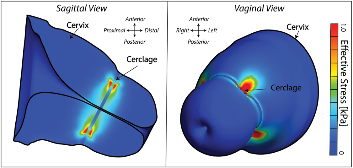
This sort of work is (pardon the pun) in its infancy. While digital twins of some organs, like the heart, are already advanced, we’re still doing the fundamental research to understand the uterus, cervix, and overall functioning of the women’s reproductive system.
It’s a big deal because these twins are only as good as the underlying data. Unlike in aerospace engineering, there’s no detailed atlas of the orientation of fibers that comprise the cervix. We don’t yet have standardized measurements of how the uterus stretches week by week across pregnancy. Shockingly, information about fundamental properties of pregnancy varies widely in the literature, if they’ve been documented at all.
That lack of information is a problem for researchers, physicians, industry, and patients. Without foundational knowledge, femtech startups are left taking guesses or burning time and capital answering basic questions. Founders regularly ask me questions about the size and shape of the uterus and cervix or how they’re packaged in the vaginal canal. These inventors are building important technologies like more effective contraception devices and treatments to prevent preterm birth. It’s vital for them to have access to high-quality information.
To support safe, effective innovation in femtech and better clinical outcomes, we need sustained investment in the full research pipeline: from curiosity-driven science to technology transfer and clinical applications. That means support from philanthropy and industry as well as from the federal government. One reason my lab has been able to continue this work over the years is the ongoing support of the Iris Fund, a foundation focused on advancing research in preterm birth and supporting families that experience high-risk pregnancies. When other sources of funding have fallen short, the Iris Fund has stepped in to fill in the gaps.
We’ve figured out how to model jet wings and SUVs — understanding how the uterus stretches through the course of pregnancy is within our grasp. But we can’t skip straight to product design. First, we need the science.
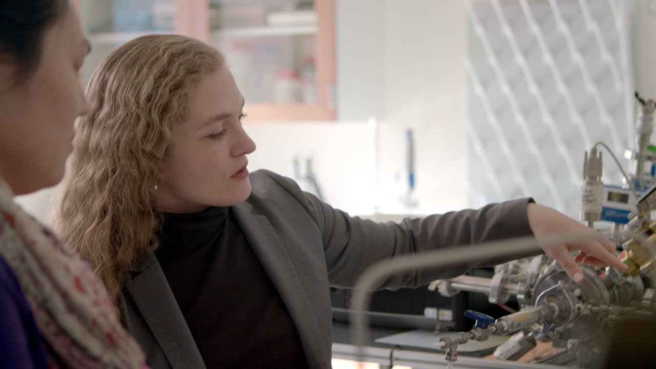
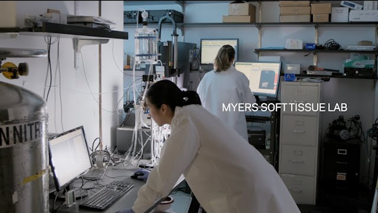
Kristin Myers is a professor of mechanical engineering, director of the Myers Soft Tissue Lab at Columbia University, and an expert in women's health engineering research.
Lead Photo Caption: Kristin Myers spoke on a panel about digital twins in medicine at the 2025 Aspen Ideas Health festival.
Lead Photo Credit: Aspen Institute