
P. James Schuck
J. Thomas "Tommy" Vaughan, Jr.
About the Study
Journal: Nature Nanotechnology
Title: “Selective targeting of visceral adiposity by polycation nanomedicine.”
Authors : Qianfen Wan (1)†, Baoding Huang (7,2)†, Tianyu Li (2), Yang Xiao (2), Ying He (1), Wen Du (3), Branden Z. Wang (1), Gregory F. Dakin (4), Michael Rosenbaum (3,5), Marcus D. Goncalves (6), Shuibing Chen (4), Kam W. Leong (2), Li Qiang (1)
- Naomi Berrie Diabetes Center and Department of Pathology and Cell Biology, Columbia University
- Department of Biomedical Engineering, Columbia University
- Department of Medicine, Columbia University
- Department of Surgery, Weill Cornell Medicine
- Department of Pediatrics, Columbia University
- Department of Medicine, Department of Surgery, Weill Cornell Medicine
- Department of Orthopedic Surgery, The Sixth Affiliated Hospital, Sun Yat-Sen University and Guangdong Provincial Key Laboratory of Orthopedics and Traumatology; Guangzhou, China
This work was supported by Russell Berrie Foundation (L.Q. and Q.W.), Blavatnik SIRS funding (L.Q. and K.W.L.), National Institutes of Health grant RO1AR073935 and the U.S. Army Medical Research grant W81XWH1910463 (K.W.L.), and The Manoogian Simone Foundation (M.D.G).
COI: A patent application is pending. All other authors declare no conflict of interest.
Journal: Biomaterials
Title: “Polycationic PAMAM ameliorates obesity-associated chronic inflammation and focal adiposity.”
Authors: Baoding Huang (1,2), Qianfen Wan (3), Tianyu Li (2), Calhoun Carmen (3), Kam W. Leong (2), and Li Qiang (3)
- Department of Orthopaedic Surgery, The Sixth Affiliated Hospital, Sun Yat-sen University and Guangdong Provincial Key Laboratory of Orthopaedics and Traumatology, Guangzhou, China
- Department of Biomedical Engineering, Columbia University
- Naomi Berrie Diabetes Center and Department of Pathology and Cell Biology, Columbia University
The study was supported by the Russell Berrie Foundation (L.Q. and Q.W.), Blavatnik SIRS funding (L.Q. and K.W.L.), National Institutes of Health grant RO1AR073935 and the U.S. Army Medical Research grant W81XWH1910463 (K.W.L.).
COI: The authors have patents pending. No other competing interests are declared.
MediSCAPE
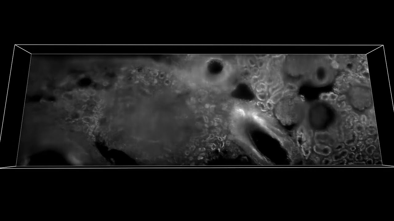
3D rendering and dynamic visualization of fresh human kidney tissue after application of topical nuclear stain (proflavine) images using MediSCAPE (2.73 mm wide strip). Sample shows signs of arterionephrosclerosis.
“Understanding whether tissues are staying healthy and getting good blood supply during surgical procedures is really important,” says Hillman. “We also realized that if we don’t have to remove (and kill) tissues to look at them, we can find many more uses for MediSCAPE, even to answer simple questions such as ‘what tissue is this?’ or to navigate around precious nerves. Both of these applications are really important for robotic and laparoscopic surgeries where surgeons are more limited in their ability to identify and interact with tissues directly.”
A critical final step for the team was to reduce the large format of the standard SCAPE microscopes in Hillman’s lab to something that would fit into an operating room and could be used by a surgeon in the human body. Post-doctoral fellow Wenxuan Liang worked with the team to develop a smaller version of the system with a better form factor, and a sterile imaging cap. PhD candidate Malte Casper helped to acquire the team’s first demonstration of MediSCAPE in a living human, collecting images of a range of tissues in and around the mouth. These results included rapidly imaging while a volunteer literally licked the end of the imaging probe, producing detailed 3D views of the papillae of the tongue.
Eager to take this technology to the next level with a larger clinical trial, the team is currently working on commercialization and FDA approval. Hillman adds, “We are just so amazed to see what MediSCAPE reveals every time we use it on a new tissue, and especially that we barely ever even needed to add dyes or stains to see structures that pathologists can recognize.”
Hillman and her team hope that MediSCAPE will make standard histology a thing of the past, putting the power of real-time histology and decision making into the surgeon’s hands.
About the Study
Journal: Nature Biomedical Engineering
Title: High-speed light-sheet microscopy for the in-situ acquisition of volumetric histological images of living tissue
Authors: Kripa B. Patel 1, Wenxuan Liang 1, Malte J. Casper 1, Venkatakaushik Voleti1, Wenze Li1, Alexis J. Yagielski1, Hanzhi T. Zhao 1, Citlali Perez-Campos 1, Joyce M. Liu1, Elizabeth Philipone2, Angela J. Yoon 2, Kenneth P. Olive3, Shana M. Coley 4 and Elizabeth M. C. Hillman 1 1Laboratory for Functional Optical Imaging, Department of Biomedical Engineering and Radiology and the Mortimer B. Zuckerman Mind Brain Behavior Institute, Columbia University 2Department of Oral and Maxillofacial Pathology, Columbia University Irving Medical Center 3Division of Digestive and Liver Disease, Herbert Irving Comprehensive Cancer Center, Columbia University Irving Medical Center 4Department of Pathology and Cell Biology, Vagelos College of Physicians and Surgeons, Columbia University Medical Center
Funding for this work was provided by the Columbia-Coulter Translational Research Partnership and the Coulter Foundation Early Career programme to E.M.C.H; the National Institutes of Health BRAIN initiative grants U01NS09429, UF1NS108213 to E.M.C.H and U19NS104649 to Costa; NCI grant U01CA236554 to E.M.C.H. and Brenner; the National Science Foundation NSF-GRFP DGE - 1644869 to K.B.P., IGERT 0801530 to V.V. and CAREER CBET-0954796 to E.M.C.H.; the Simons Foundation Collaboration on the Global Brain 542951 to E.M.C.H.; the Department of Defense MURI W911NF-12-1-0594 to E.M.C.H.; and the Kavli Institute for Brain Science to E.M.C.H.
COI: Intellectual property related to SCAPE microscopy is held by Columbia University and is licensed to Leica Microsystems for certain applications. The authors of this study could benefit financially from commercial development of this technology.
About the Study
Journal: Nature Biotechnology
Title: A programmable encapsulation system improves delivery of therapeutic bacteria in mice
Authors: Tetsuhiro Harimoto 1,6, Jaeseung Hahn 1,6, Yu-Yu Chen 1, Jongwon Im 1, Joanna Zhang 1, Nicholas Hou 1, Fangda Li 2, Courtney Coker 1, Kelsey Gray 1, Nicole Harr 1, Sreyan Chowdhury 1,2, Kelly Pu 1, Clare Nimura 1, Nicholas Arpaia 2,3, Kam Leong 1,4, and Tal Danino 1,3,5
- Department of Biomedical Engineering, Columbia Engineering
- Department of Microbiology and Immunology, Vagelos College of Physicians and Surgeons, Columbia University
- Herbert Irving Comprehensive Cancer Center, Columbia University
- Department of Systems Biology, Columbia University Medical Center
- Data Science Institute, Columbia University
This work was supported by the DoD LC160314 (T.D.), DoD BC160541 (T.D.), NIH R01CA249160 (T.D.), NIH U01CA247573 (T.D.), NIH F99CA253756 (T.H.), and Honjo International Foundation Scholarship (T.H.).
COI: T.H., J.H., K.L. and T.D. have filed a provisional patent application with the US Patent and Trademark Office related to this work (01001/009982).
Header image: Probiotic bacteria (E. coli Nissle 1917 strain, green) is engineered to controllably evade immune system (macrophage, transparent) using a genetically encoded encapsulation system (capsular polysaccharides with circuit board, shown as transparent coating surrounding the bacterial cells). This system was used to enhance delivery of therapeutic bacteria for cancer therapy. (Ella Marushchenko, Alex Tokarev, Danino Lab/Columbia Engineering)
About the Study
Journal: Communications Biology, an open-access journal from Nature Portfolio
Title: Combination of antiviral drugs inhibits SARS-CoV2 polymerase and exonuclease and demonstrates COVID-19 therapeutic potential in viral cell culture
Authors: Xuanting Wang1,2,11, Carolina Q. Sacramento3,4,11, Steffen Jockusch1,5,11, Otavio Augusto Chaves3,4, Chuanjuan Tao1,2, Natalia Fintelman-Rodrigues3,4, Minchen Chien1,2, Jairo R. Temerozo6,7, Xiaoxu Li1,2, Shiv Kumar1,2, Wei Xie8, Dinshaw J. Patel8, Cindy Meyer9, Aitor Garzia9, Thomas Tuschl9, Patricia T. Bozza3, James J. Russo1,2, Thiago Moreno L. Souza3,4 & Jingyue Ju1,2,10
- Center for Genome Technology and Biomolecular Engineering, Columbia University
- Department of Chemical Engineering, Columbia University
- Laboratory of Immunopharmacology, Oswaldo Cruz Institute (IOC), Oswaldo Cruz Foundation (Fiocruz), Rio de Janeiro
- National Institute for Science and Technology for Innovation on Diseases of Neglected Population (INCT/IDN), Center for Technological Development in Health (CDTS), Oswaldo Cruz Foundation (Fiocruz), Rio de Janeiro
- Department of Chemistry, Columbia University
- Laboratory on Thymus Research, Oswaldo Cruz Institute (IOC), Oswaldo Cruz Foundation (Fiocruz), Rio de Janeiro
- National Institute for Science and Technology on Neuroimmunomodulation (INCT/NIM), Oswaldo Cruz Institute (IOC), Oswaldo Cruz Foundation (Fiocruz), Rio de Janeiro
- Laboratory of Structural Biology, Memorial Sloan-Kettering Cancer Center
- Laboratory of RNA Molecular Biology, Rockefeller University
- Department of Molecular Pharmacology and Therapeutics, Columbia University
This work was supported by the Jack Ma Foundation, a gift from Columbia Engineering Member of the Board of Visitors Dr. Bing Zhao, and Fast Grants (to Jingyue Ju), the Maloris Foundation and the Memorial Sloan- Kettering Core Grant (P30CA008748) (to Dinshaw J. Patel), a grant from The JPB Foundation to Rockefeller University (to Thomas Tuschl). Funding was also provided by Conselho Nacional de Desenvolvimento Cientifico e Tecnologico (CNPq, 441019/2020-0, 307162/2017-6), Fundacao de Amparo a Pesquisa do Estado do Rio de Janeiro (FAPERJ, E-26/210.182/2020, E-26/201.067/2021, E-26/210.112/2020) and Coordenacao de Aperfeicoamento de Pessoal de Ni vel Superior -Brasil (CAPES) - Finance Code 001 (to Thiago Moreno L. Souza and Patricia T. Bozza). CNPq, CAPES and FAPERJ also support the National Institutes of Science and Tech-nology Program (INCT-IDPN, 465313/2014-0). Oswaldo Cruz Foundation/FIOCRUZ supports this study under the auspices of the Inova Program (B3-Bovespa funding, VGPDI-032-ARVC-20) (to Thiago Moreno L. Souza).
COI: The authors declare no competing interests.
About the Studies
Journal: Science Advances
Title: Ionic communication for implantable bioelectronics
Authors: Zifang Zhao, George Spyropoulos, Claudia Cea, Jennifer N. Gelinas and Dion Khodagholy.
Jurnal: Advanced Science
Title: Anisotropic Ion Conducting Particulate Composites for Bioelectronics
Authors: Dickson R. Yao, Han Yu, Onni J. Rauhala, Claudia Cea, Zifang Zhao, Jennifer N. Gelinas and Dion Khodagholy.
Department of Electrical Engineering.
Department of Neurology.
Institute for Genomic Medicine.
Both these studies were supported by the Columbia University School of Engineering and Applied Science, the Columbia University Medical Center, Department of Neurology and Institute for Genomic Medicine, NIH grant 1U01NS108923-01, NIH grant R01NS118091, NIH grant R21 EY 32381-01, NSF CAREER award 1944415, and NSF EAGER grant 2027135.
COI: The authors declare no financial or other conflicts of interest.