Acceleration Funds for 2022-2024
Project 1: Hemodynamic Predictors of Aortic Root Aneurysm Progression Using Computational Modeling
Abstract
Global rates of preterm birth (PTB) remain astonishingly high, accounting for the majority of infant deaths. Our multidisciplinary team seeks to understand which physiological factors support a healthy pregnancy by leveraging machine learning to test hypotheses about the mechanical environment of pregnancy. Currently, risk assessment and management of spontaneous PTB rely heavily on sonographer assessment of cervical length (CL), an incomplete measure of cervical structural health. Although routine CL screening has identified some high-risk patients, it is not universally adopted because predictions remain unreliable. It is known that age, race, and surgical history are also associated with spontaneous PTB. Our past computational biomechanical models show the cervical function depends on its volume and shape more than cervical length alone. Based on this premise, our team of engineers, radiologists, biostatisticians, and clinicians propose to collect electronic medical record (EMR) data and clinical cervical ultrasound (US) images using an IRB-approved protocol and to use machine learning to develop and optimize PTB prediction models. Using the US images, we will train a novel deep-learning algorithm to extract cervical size and shape automatically (Aim 1). Then we will combine these raw images and cervical structural features with EMR data to build a high-dimensional, patient-specific PTB prediction model (Aim 2). In future work, these data will be leveraged in biomechanical simulations to gain a mechanistic understanding of a healthy, term delivery.
Principal Investigators
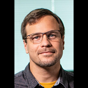
Vijay Vedula is an assistant professor of mechanical engineering at Columbia University. He received his Bachelor’s and Master’s degrees from India, a PhD from Johns Hopkins University, and did his postdoctoral work at Stanford University through a fellowship from the Child Health Research Institute (CHRI) at Stanford. His expertise is in computational modeling of cardiovascular biomechanics in disease and development. Dr. Vedula’s research group focuses on developing novel computational methods for personalized modeling of cardiac biomechanics including blood flow, tissue response, and valvular interactions. These tools are then applied to both congenital and adult cardiac disease, with emphasis on understanding disease progression and surgical planning.
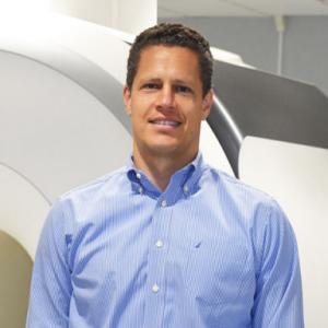
Dr. Hiroo Takayama is Chief of Adult Cardiac Surgery at NewYork-Presbyterian/Columbia University Irving Medical Center. Dr. Takayama attended University of Tokyo for Medical School, did his internship at University of Tokyo, general surgery residency in University of Washington Medical Center, and cardio-thoracic surgery fellowship in New-York Presbyterian/Columbia University Medical Center. He has more than 20 years of experience in treating patients with heart and vascular problems. As the Director of the Aortic Surgery Program and co-Director of the Aortic Center, the Hypertrophic Cardiomyopathy Center, and the Marfan Clinic at NYP/Columbia, Dr. Takayama has contributed to the care of over 4,000 patients by performing a variety of aortic and cardiac surgical procedures, approximately 300-350 operations every year. Dr. Takayama is internationally recognized for his work on complex aortic surgery (minimally invasive aortic surgery, valve-sparing aortic root replacement, aortic valve repair, aortic arch and thoracoabdominal aneurysm repair), hybrid aortic surgery (thoracic endovascular aneurysm repair), myectomy for obstructive hypertrophic cardiomyopathy, and heart failure surgery. He was named one of New York Magazine’s Top Doctors in 2016 and won the 2012 Thoracic Surgery Residents Association Dwight C. McGoon Award.
Project 2: Stem cell model of embryo implantation and placenta development: discovering molecular mechanisms and advancing early pregnancy outcomes.
Abstract
The correct placenta development during embryo implantation into the uterus is critical to establishing a successful pregnancy. Disruptions in this process are the leading cause of recurrent implantation failure, miscarriages, and a host of other obstetric disorders. Pregnancy-related maternal death has more than doubled since 1987 and populations that have been historically oppressed—Black and Indigenous women—are disproportionately affected. Despite these devastating outcomes, we know very little about how the early human placenta develops, leaving treatment options for pregnancy disorders very limited. We owe this knowledge gap to the very nature of mammalian embryogenesis: once the embryo implants into the uterus, it becomes obscured from view making it challenging to glean mechanistic knowledge about this process. We propose to combine stem cell and patient-derived organoids, mechanically tunable biomaterials, and CRISPR gene editing, to create the first organoid platform that models nascent placenta development in contact with uterine tissues. Unlike any available experimental system, our platform will promote a much more realistic implantation microenvironment. It will enable the correct placenta-uterine signaling feedback critical for the discovery of underlying molecular mechanisms. Paired with single cell RNA sequencing, we will generate the first comprehensive atlas of implantation and elucidate the hierarchy of cell fate decisions in the making of the early placenta. Given the high-risk high-reward nature of our proposal, we apply for SIRS Fund for Engineering Innovations in Health to first develop and fully characterize our experimental platform. In the long run, our interdisciplinary approach will enable predictive insights that can be used to develop clinical screening methods to advance pregnancy health.
Principal Investigators
Mijo Simunovic is an assistant professor of chemical engineering at Columbia University. Simunovic received their BS and MS in chemistry from the University of Zagreb. They received their first PhD in theoretical chemistry from the University of Chicago and their second PhD in condensed matter physics from the Curie Institute and the University of Paris VII. They conducted their postdoctoral research in stem cell and developmental biology at the Rockefeller University as a Junior Fellow of the Simons Society of Fellows. Their research focuses on the creation of quantitative models of embryogenesis using human pluripotent stem cells, to study the molecular mechanisms and the biomechanics of embryo implantation, gastrulation, and organ formation. Throughout their career, Simunovic has been recognized with numerous research and teaching awards, including the 2017 AAAS/Science and SciLifeLab Prize for Young Scientists in Cell and Molecular Biology and the 2021 NIH Director's New Innovator Award.
Zev Williams, MD, PhD is the Wendy D. Havens Associate Professor of Women's Health and the Chief of the Division of Reproductive Endocrinology and Infertility at Columbia University Irving Medical Center. He completed his MD and PhD training in Molecular Biology and Biochemistry at the Mount Sinai School of Medicine before continuing to the Brigham and Women’s Hospital/Massachusetts General Hospital for his residency in Obstetrics and Gynecology. After completing his fellowship in Reproductive Endocrinology and Infertility at Weill-Cornell, Dr. Williams then did a post-doctoral fellowship on RNA biology in the laboratory of Dr. Thomas Tuschl at Rockefeller University. As a physician scientist, Dr. Williams' focus has been on helping those suffering from recurrent pregnancy loss and infertility and developing novel technologies and treatments to improve patient success.
Project 3: Measuring ferroptosis in vivo using magnetic resonance spectroscopy
Abstract
Ferroptosis is a non-apoptotic form of cell death that results from the failure to detoxify lipid reactive oxygen species (ROS). Lipid radicals can react with other lipids in a self-propagating reaction within lipid membranes leading to a cascade of lipid peroxidation and membrane rupture. To date, this process has been almost entirely characterized in vitro, where ferroptosis is defined as an oxidative, iron dependent form of non-apoptotic cell death. The Olive lab previously demonstrated that tumor-selective ferroptosis can be induced in genetically engineered models of pancreatic ductal adenocarcinoma (PDAC) through the depletion of cysteine, the primary cellular source of thiol species. A major ongoing challenge to the field of ferroptosis research is the lack of a proven biomarker for ferroptosis in vivo. We propose to use magnetic resonance spectroscopy (MRS) to measure two metabolic consequences of ferroptosis initiation: lowered levels of the cysteine-derivative antioxidant glutathione (GSH) and increased lipid droplet formation. Cysteine depletion rapidly leads to loss of its labile derivative GSH. Lipid droplets form as an indirect consequence of lipid oxidative stress. We will develop a sensitive and robust MRS detection method for GSH using Semi-LASER, an analysis technique pioneered by Dr. Juchem. We will then compare the performance of this approach to an established method for detecting choline species in PDAC-bearing mice undergoing cysteine depletion. The success of this proposal will provide a critical new capability for the entire ferroptosis field that will be deployed in the Oncology Precision Therapeutics and Imaging Core (OPTIC) for Columbia users and that is readily translated to a clinical setting.
Principal Investigators
Christoph Juchem is an associate professor in the Departments of Biomedical Engineering and Radiology at Columbia University. He holds a PhD from the University of Bonn, Germany and did doctoral studies at the Max-Planck Institute for Biological Cybernetics. In his research, he develops novel magnetic resonance methods and technology to establish optimized tools for neuroscientific and clinical applications. His long-term goal as a physicist is to realize the full potential of magnetic resonance spectroscopy (MRS) as a diagnostic tool. His clinical long-term goal is to understand the role that neurochemicals play in the protection of the human central nervous system (CNS) or, alternatively, how dysfunction promotes vulnerability towards neurodegenerative and neuro-immunological diseases. He served as Co-Director of Yale’s 7T Brain MR Spectroscopy Core, Chair of the ISMRM Engineering Study group and member of the editorial board of NMR in Biomedicine. He is Vice-Chair of the ISMRM MR Spectroscopy Study Group, to be Chair in 2023.
Kenneth P. Olive is an associate professor in the department of medicine and director of the Oncology Precision Therapeutics and Imaging Core (OPTIC) Shared Resource. He holds a BS in biology from Bucknell University and a PhD in biology from M.I.T. Dr. Olive's research is dedicated to finding a cure for pancreatic cancer. The Olive laboratory performs preclinical therapeutics trials using advanced genetically engineered mouse models of pancreatic cancer. Ultimately, successful therapies will be translated into the clinical setting through our collaborations with the Pancreas Center of Columbia University. Complementing his laboratory research, Olive has also built a large-scale translational core facility called the Oncology Precision Therapeutics and Imaging Core (OPTIC). He won the Lustgarten Foundation Translational Innovator Award in 2011.