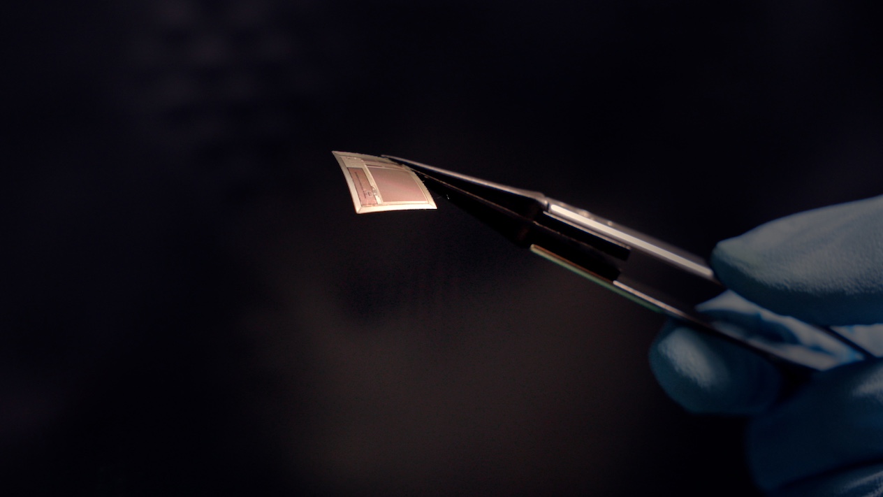
Described in a study published Dec. 8 in Nature Electronics, BISC includes a single-chip implant, a wearable “relay station,” and the custom software required to operate the system. “Most implantable systems are built around a canister of electronics that occupies enormous volumes of space inside the body,” says Ken Shepard, Lau Family Professor of Electrical Engineering, professor of biomedical engineering, and professor of neurological sciences at Columbia University, who is one of the senior authors on the work and guided the engineering efforts. “Our implant is a single integrated circuit chip that is so thin that it can slide into the space between the brain and the skull, resting on the brain like a piece of wet tissue paper.” 

Shepard was joined in the BISC effort by senior and co-corresponding author Andreas S. Tolias, PhD, professor at the Byers Eye Institute at Stanford University and co-founding director of the Enigma Project. Tolias’s pioneering work training AI models on large-scale neural datasets — including datasets recorded in the Tolias laboratory using BISC — enabled the team to evaluate the device’s neural decoding performance. “BISC turns the cortical surface into an effective portal, delivering high-bandwidth, minimally invasive read–write communication with AI and external devices,” Tolias says. “Its single-chip scalability paves the way for adaptive neuroprosthetics and brain-AI interfaces to treat many neuropsychiatric disorders, such as epilepsy.”

Dr. Brett Youngerman, assistant professor of neurological surgery at Columbia University and a neurosurgeon at NewYork-Presbyterian/Columbia University Irving Medical Center, served as the chief clinical collaborator on the project. “This high-resolution, high-data-throughput device has the potential to revolutionize the management of neurological conditions from epilepsy to paralysis,” he says. Youngerman, Shepard, and NewYork-Presbyterian/Columbia epilepsy neurologist Dr. Catherine Schevon were recently awarded a grant from the National Institutes of Health to implement BISC in the management of drug-resistant epilepsy. “The key to effective brain-computer interface devices is to maximize the information flow to and from the brain, while making the device as minimally invasive in its surgical implantation as possible. BISC surpasses previous technology on both fronts,” continues Youngerman.

“Semiconductor technology has made this possible, allowing the computing power of room-sized computers to now fit in your pocket,” Shepard says. “We are now doing the same for medical implantables, allowing complex electronics to exist in the body while taking up almost no space.”

Smaller, Safer, and Faster

BCIs work by interfacing with the electrical signals that neurons use to transfer information throughout the brain. Today’s state-of-the-art BCIs, used in medical contexts, are constructed from individual microelectronic components, including amplifiers, data converters, radio transmitters, and power management circuits. To accommodate all these devices, a large canister of electronics must be surgically implanted in the body, either by removing a portion of the skull or by placing the device in another location, such as the chest, and running wires to the brain.

BISC works differently. The entire implant, which occupies less than 1/1000th the size of a conventional device, is a single complementary metal-oxide-semiconductor (CMOS) integrated circuit chip thinned to just 50 μm. With a total volume of approximately 3 mm³, the flexible chip conforms to the surface of the brain. This micro-electrocorticography (µECoG) device integrates 65,536 electrodes, 1,024 simultaneous recording channels, and 16,384 stimulation channels. By leveraging the large-scale manufacturing techniques developed in the semiconductor industry, these implants can be easily manufactured at scale.

The single-chip implant includes a radio transceiver, wireless powering circuit, digital control, power management, data conversion, and the analog circuits required to support the recording and stimulation interfaces. The battery-powered relay station powers and communicates with the implant, transferring data via a custom ultrawideband radio link that achieves 100 Mbps data bandwidths — a connection with at least 100 times higher throughput than any competing wireless BCI device. The relay station is itself an 802.11 WiFi device, in effect forming a relayed wireless network connection from any computer to the brain. 

BISC has its own instruction set, supported by an extensive software stack, which together constitute a computing architecture designed for BCIs. As demonstrated in this study, these high-bandwidth recording capabilities allow brain-signal patterns to be submitted to advanced machine-learning or deep-learning frameworks for decoding complex intentions, perceptions, or states.

 “By integrating everything on one piece of silicon, we’ve shown how brain interfaces can become smaller, safer, and dramatically more powerful,” Shepard says.

The BISC implant was manufactured using TSMC’s versatile 0.13-μm Bipolar-CMOS-DMOS (BCD) technology. This manufacturing process integrates three technologies onto a single chip to create mixed-signal integrated circuits (ICs). This integration enables the efficient combination of digital logic (from CMOS), high-current and high-voltage analog functions (from bipolar and DMOS transistors), and power devices (from DMOS), all of which are essential for BISC.

From Lab to Clinic

To make this technology available to doctors and patients, Shepard’s group partnered closely with Youngerman at NewYork-Presbyterian/Columbia University Irving Medical Center. Together, they refined surgical methods to safely implant the paper-thin device in a preclinical model and demonstrated its recording quality and stability, as described in the current study. Studies in human patients for short-term intraoperative recordings are underway.

“These initial studies give us invaluable data about how the device performs in a real surgical setting,” Youngerman says. “The implants can be inserted through a minimally invasive incision in the skull and slid directly onto the surface of the brain in the subdural space. The paper-thin form factor and lack of brain-penetrating electrodes or wires tethering the implant to the skull minimize tissue reactivity and signal degradation over time.”

Extensive pre-clinical testing of BISC in the motor and visual cortices drew on collaborations with both Dr. Tolias and Bijan Pesaran, professor of neurosurgery at the University of Pennsylvania, both of whom are leaders in computational and systems neuroscience.

“The extreme miniaturization by BISC is very exciting as a platform for new generations of implantable technologies that also interface with the brain with other modalities such as light and sound,” Pesaran says.

Developed under the Neural Engineering System Design program of the Defense Advanced Research Projects Agency (DARPA), BISC combines Columbia’s strengths in microelectronics, Stanford’s and Penn’s cutting-edge neuroscience, and NewYork-Presbyterian/Columbia University Irving Medical Center’s surgical innovation.

Toward Real-World Applications

To accelerate translation, the Columbia and Stanford teams launched Kampto Neurotech, a spin-off company founded by Columbia electrical engineering alumnus Dr. Nanyu Zeng, one of the project’s lead engineers. Kampto Neurotech is developing commercial versions of the chip for preclinical research applications and raising funds to advance the system toward human use.

“This is a fundamentally different way of building BCI devices,” Zeng says. “In this way, BISC has technological capabilities that exceed those of competing devices by many orders of magnitude.” 

In a technological landscape driven by advances in artificial intelligence, BCI technologies have drawn considerable recent interest in both restoring function to those affected by neurological conditions and in potentially augmenting human capabilities by providing direct interfaces to the brain.

“By combining ultra-high resolution neural recording with fully wireless operation, and pairing that with advanced decoding and stimulation algorithms, we are moving toward a future where the brain and AI systems can interact seamlessly — not just for research, but for human benefit,” says Shepard. “This could change how we treat brain disorders, how we interface with machines, and ultimately how humans engage with AI.”


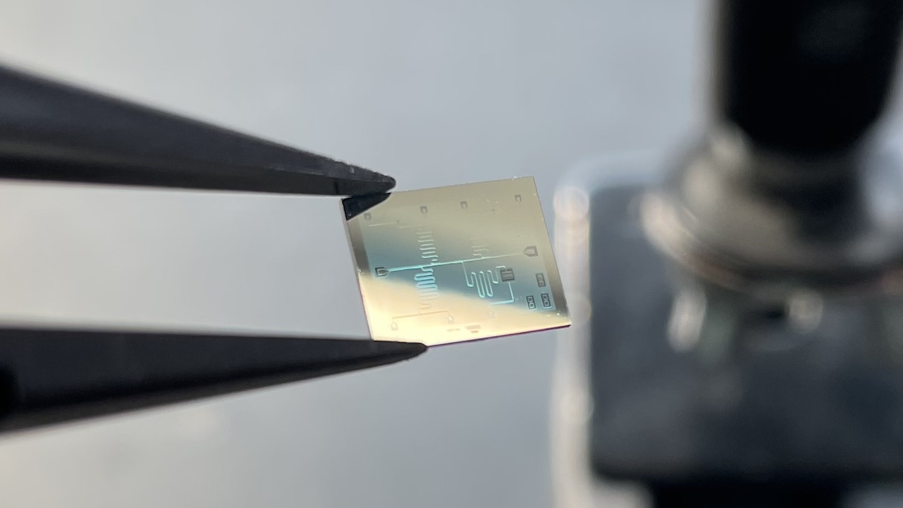
Lead Photo Caption: The BISC implant shown here is roughly as thick as a human hair. 

Lead Photo Credit: Columbia Engineering

About The Study

Journal: Nature Electronics

DOI: 10.1038/s41928-025-01509-9

Title: Stable, chronic in-vivo recordings from a fully wireless subdural-contained 65,536-electrode brain-computer interface device

Authors: Taesung Jung, Nanyu Zeng, Jason D. Fabbri, Guy Eichler, Zhe Li, Erfan Zabeh, Anup Das, Konstantin Willeke, Katie E. Wingel, Agrita Dubey, Rizwan Huq, Mohit Sharma, Yaoxing Hu, Girish Ramakrishnan, Kevin Tien, Paolo Mantovani, Abhinav Parihar, Heyu Yin, Denise Oswalt, Alexander Misdorp, Ilke Uguz, Tori Shinn, Gabrielle J. Rodriguez, Cate Nealley, Sophia Sanborn, Ian Gonzales, Michael Roukes, Jeffrey Knecht, Daniel Yoshor, Peter Canoll, Eleonora Spinazzi, Luca P. Carloni, Bijan Pesaran, Saumil Patel, Joshua Jacobs, Brett Youngerman, R. James Cotton, Andreas Tolias, Kenneth L. Shepard

Funding/Acknowledgments: This work was partly supported by the Defense Advanced Research Projects Agency (DARPA) under Contract N66001-17-C-4001, the Department of the Defense Congressionally Directed Medical Research Program under Contract HT9425-23-1-0758, the National Science Foundation under Grant 1546296, and the National Institutes of Health under Grant R01DC01949

This year’s winners all received electronic prizes. Find out more information about the MakeCU submissions here


Lead Photo Caption: First place-winning team, Dormmate. The team’s smart doorbell system allows you to remotely unlock your door, even when you’re not home.

Lead Photo Credit: Columbia University Robotics

From flat sheets to complex structures

One major way that developing embryos build their organs is through furrowing — that is, they form pockets in tissues, which eventually become the sites of folds. "Just as a flat sheet of paper can be folded into a crane, a flat embryonic tissue can be folded into the precursor of an organ," said Andrew Countryman, a doctoral student in biomedical engineering at Columbia and the study's first author.

Previous research has developed many tools for manipulating the proteins and other molecules that direct how cells behave. However, scientists lacked similar techniques for systematically controlling the mechanical forces that ultimately shape embryos. 

In the new study, Kasza, Countryman, and their colleagues experimented with the fruit fly, a common lab animal. "As developmental processes and machinery are highly conserved across animals, these findings in fruit flies provide insight into development in all animals, including humans," Countryman said.

Light-sensitive tools built with CRISPR

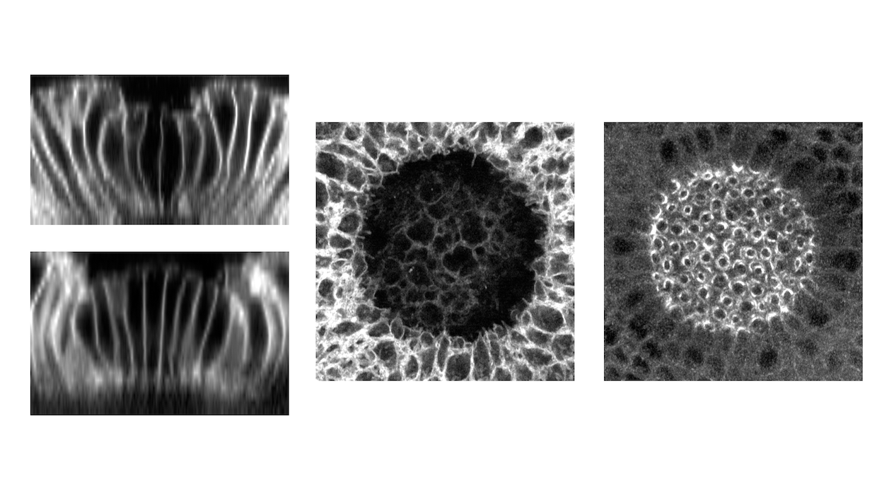
The researchers tinkered with proteins that cells use to generate mechanical forces, making these molecules responsive to light. By shining patterns of specific wavelengths of light on fruit fly embryos genetically modified to produce these proteins, they could in turn control patterns of forces during their development. 

The new study used the gene-editing system CRISPR-Cas9 to add a light-sensitive module to genes that naturally exist in fruit flies. The resulting molecules are the first tools that let scientists use light to control an animal's own genes to direct mechanical forces in live embryos. They are also the first tools that enable scientists to employ light to control cell-generated forces in a tunable way, instead of just switching such forces on and off, Countryman said.

The researchers specifically modified proteins that help cells contract, one method by which tissues can generate furrows. The resulting tools, called endogenous OptoRhoGEFs, helped the scientists discover that the depth of a furrow depends on the amount of these contraction-linked proteins that get summoned to a cell's membrane. They also found that stiff layers of proteins within embryos could dramatically influence the ways in which tissues furrowed.

Implications for human health

"Similarly to fly embryos, human embryos extensively employ furrowing processes during development," Countryman said. "A failure of tissues to furrow properly is associated with common and devastating congenital disorders, such as spina bifida. Improved understanding of developmental processes will help identify and treat these conditions."

This new technique may one day help scientists better analyze tissue and organ development and disease, using light to fold basic sheets of cells into complex 3D structures in the lab instead of the more complex environments inside living animals, Countryman said.

In addition, "small, controllable, cell-based machines have promising use in medical contexts, where they can serve as biocompatible probes during medical procedures," he added. "They could also be used as small, aqueous, remotely pilotable vehicles to explore and survey new environments."

In the future, the researchers hope to use their new strategy to examine other ways in which tissues furrow, as well as tissue behaviors other than furrowing, such as bending, stretching, and flowing. "These basic modes of tissue deformation are used in different combinations and sequences to build a wide variety of tissues, organs, and body forms," Countryman said.


Lead Photo Caption: Re-engineering force-regulating proteins inside cells to control their behavior with specific wavelengths of light. Side view of a small group of optogenetically activated cells forming a furrow toward the inside of the embryo (top image, far left); Side view of a large group of optogenetically activated cells bending toward the outside of the embryo (bottom image, far left); Top view of a large group of optogenetically activated cells bending toward the outside of the embryo (center); Patterns of myosin–a contractile protein–associated with optogenetic activation of a large group of cells (far right). 

Lead Photo Credit: Andrew Countryman/Kasza lab

About The Study

Journal: Nature Communications

Title: Endogenous OptoRhoGEFs reveal biophysical principles of epithelial tissue furrowing

DOI: 10.1038/s41467-025-62483-6

Authors: Andrew D. Countryman, Caroline A. Doherty, R. Marisol Herrera-Perez, Karen E. Kasza.

Funding/Acknowledgements: The researchers thank Stas Shvartsman and Liz Gavis for their contributions to conceptualization of the endogenous optogenetic tools and for helpful discussions. They thank Bex Pendrak, Sameer Thukral, and Kasza Lab members for helpful discussions. Stocks obtained from the Bloomington Drosophila Stock Center (NIH P40OD018537) were used in this study. This work was performed in part at the Live Imaging and Bioenergetics Facility at the Advanced Science Research Center at The Graduate Center of the City University of New York. This work was supported by NIH Grant R35GM138380 to Karen E. Kasza and NIH Grant 1F31HD118793-01 to Andrew D. Countryman. Karen E. Kasza holds an NSF CAREER Award, Packard Fellowship, and Sloan Research Fellowship in Physics.

The authors declare no competing interests.

Subscribe to Press Release